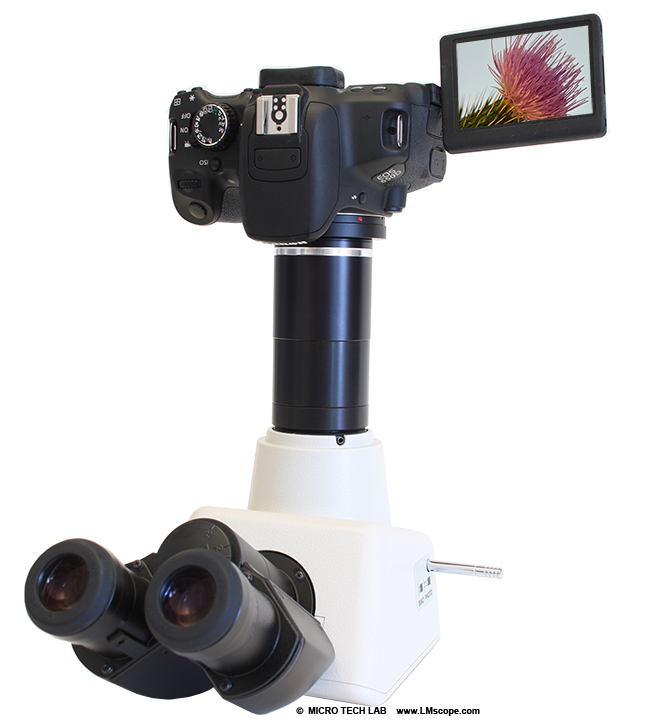
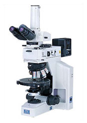
An optical microscope equipped with five objectives and Deltapix Invenio digital camera. The microscope can be used in differential interference contrast (DIC) imaging, which is an imaging method based on the contrast difference of the samples. With DIC it is possible to see details from optically transparent samples which are invisible in the ordinary microscope images.
- Nikon Eclipse Me600 Manual Instructions
- Nikon Eclipse Me600 Microscope Manual
- Nikon Eclipse Me600 Manual Troubleshooting
Nikon 1 J3 使用説明書 (5.15 MB) Nikon 1 J2 使用説明書 (3.74 MB) Nikon 1 V1 使用説明書 (3.32 MB) Nikon 1 J1 使用説明書 (3.18 MB) Nikon 1 V1/ Nikon 1 J1 追加機能のご案内 (483 KB) Nikon 1 S1 使用説明書 (4.81 MB). View and Download Nikon Eclipse E600 instructions manual online. Eclipse E600 microscope pdf manual download. Materials and Device Characterization We offer access to a wide range of characterization techniques for materials and devices; optic microscopy, high resolution scanning and transmission electron microscopy, x-ray diffraction, scanning probe techniques and a variety of electric, optic and magnetic characterization of materials, structures and devices. Vocabulary Quiz Answers Nikon Eclipse E-200 Microscope - University of Arizona Kindle File Format Jenapol Manual Honda Atv Owners Manuals Online Nikon Eclipse Me600 Manual - peugeotocm.com Alcatel 4400 User Manual - pompahydrauliczna.eu Nikon Coolscan Repair Manual - orrisrestaurant.com Hp Officejet Manuals.
- Optics: 5X/0.15, 10X/0.30, 20X/0.46, 50X/0.80, 100X/0.90
- Option for differential interference contrast (DIC) imaging


Last updated: 8.1.2013
Nikon Eclipse Me600 Manual Instructions
Description:
The SUSS PM5 is a probing station for electrical (DC and HF) measurements on wafers, chips and substrates up to 150 mm. It has four SUSS ProbeHeads placed on the stable platen, with space for additional probes if necessary. The platen height can be adjusted up to 40 mm allowing a quick and easy setup of the system. The additional contact separation of 200 μm ensures accurate fine adjustment of the probe platen. The 4 ProbeHeads (X, Y, Z movements) are connected to the platen by vacuum. They can be equipped with tungsten needles, with radius down to 2µm. A metal bridge, which spans the width of the probe system and is fastened to the base, support the microscope. This is equipped with ExtraLongWorkingDistance 2x, 10x, 20x objectives and 10x ocular. It has a third ocular for camera imaging (not installed). The chuck stage can be X, Y and Z moved and can be heated/cooled. All knobs are located to allow easy and precise movement of the chuck stage with just one hand. The X and Y axes can be adjusted independently. Once it has reached the test position, the stage locks into place and provides additional fine adjustment in the Z direction. A pull-out stage permits quick and ergonomic loading and unloading of your DUT.Working principle

Nikon Eclipse Me600 Microscope Manual
First place the DUT on the chuck and activate the vacuum for a tight fixing. Switch-on the light for the microscope. Operate X,Y,Z movements of the chuck, of the platen and of the ProbeHead to contact the needles to the DUT pads. The needles are in electrical contact with the ProbeHead arm and to the BNC cables for electrical measurements with external electronic instrumentation. Up to 4 contact points can be operated independently at the same time thanks to the 4 ProbeHaeds. Remember to switch-off microscope light when measuring.
Nikon Eclipse Me600 Manual Troubleshooting
Specifications
SÜSS MicroTec Manual Electric Probe System PM5 specifications